Home

cél Diszkriminatív Hölgyem ni sem Érik Tábornok Uralom

Nickel-based superalloy microstructure characterized by EBSD | Bruker

Powder Morphology & Cross-sectional Scanning Electron Microscopy (SEM)  Images of Commercially Available Metal Powders | PPT
Powder Morphology & Cross-sectional Scanning Electron Microscopy (SEM) Images of Commercially Available Metal Powders | PPT

SEM image of nickel particles on coated carbon paper. Ni particle sizes...  | Download Scientific Diagram
SEM image of nickel particles on coated carbon paper. Ni particle sizes... | Download Scientific Diagram

Scanning electron microscope (SEM) of Ni–Co precursor, a, b reaction... |  Download Scientific Diagram
Scanning electron microscope (SEM) of Ni–Co precursor, a, b reaction... | Download Scientific Diagram
SEM images of (A and B) the as-obtained Ni foam at different... | Download  Scientific Diagram
SEM images of (A and B) the as-obtained Ni foam at different... | Download Scientific Diagram

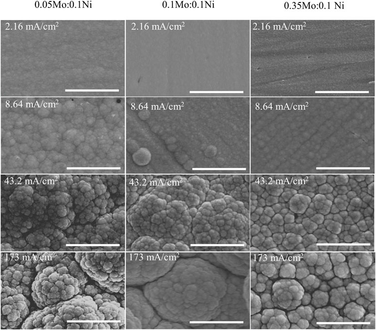
Frontiers | Effect of solution pH, precursor ratio, agitation and  temperature on Ni-Mo and Ni-Mo-O electrodeposits from ammonium citrate baths
Frontiers | Effect of solution pH, precursor ratio, agitation and temperature on Ni-Mo and Ni-Mo-O electrodeposits from ammonium citrate baths

a) SEM image of the 3D structure of nickel foam, (b – d) di ff erent... |  Download Scientific Diagram
a) SEM image of the 3D structure of nickel foam, (b – d) di ff erent... | Download Scientific Diagram
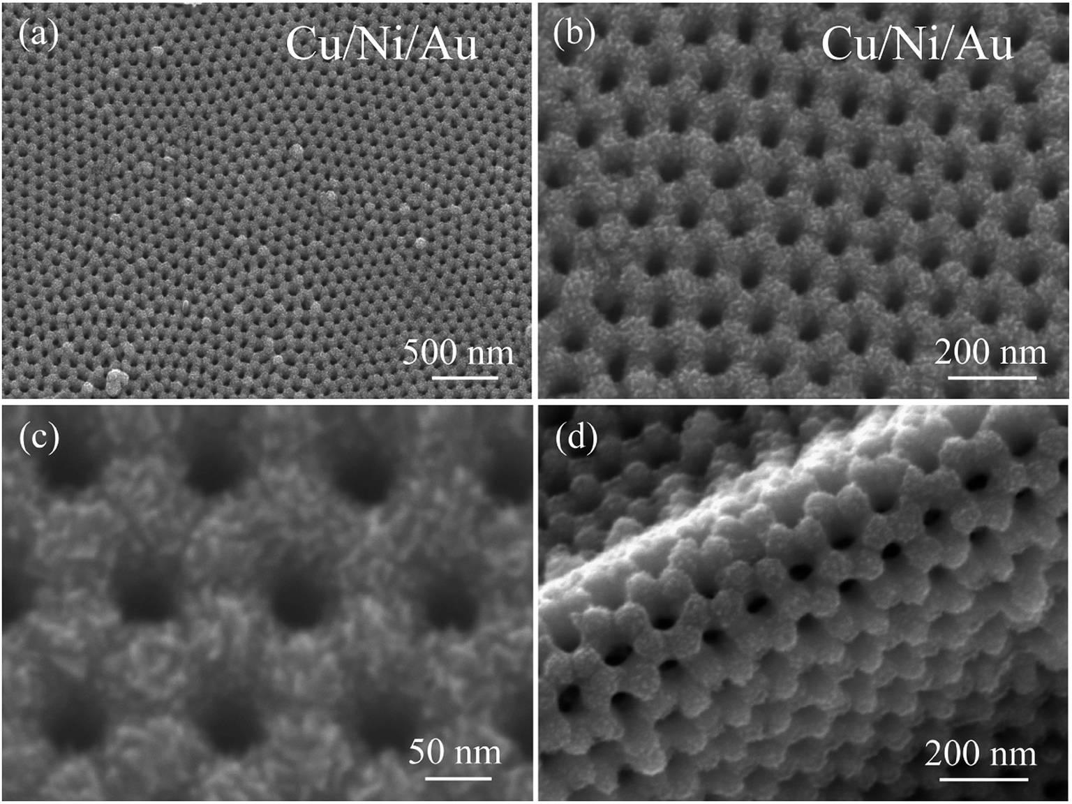
Synthesis of an ordered nanoporous Cu/Ni/Au film for sensitive  non-enzymatic glucose sensing - RSC Advances (RSC Publishing)  DOI:10.1039/D0RA01224F
Synthesis of an ordered nanoporous Cu/Ni/Au film for sensitive non-enzymatic glucose sensing - RSC Advances (RSC Publishing) DOI:10.1039/D0RA01224F

Figure S1. SEM images of plain nickel foam. | Download Scientific Diagram

SEM images. Ni nanoparticles formed at (a) 240°C, (b) 255°C, (c) 270°C,...  | Download Scientific Diagram
SEM images. Ni nanoparticles formed at (a) 240°C, (b) 255°C, (c) 270°C,... | Download Scientific Diagram

SEM images of (a) bare Ni foam, (b) Ni 3 S 2 /NF and (c, d) Fe-Ni 3 S 2...  | Download Scientific Diagram
SEM images of (a) bare Ni foam, (b) Ni 3 S 2 /NF and (c, d) Fe-Ni 3 S 2... | Download Scientific Diagram
SEM image of foam Ni. | Download Scientific Diagram

SEM images of Ni nanowires under different magnification (a,c) and... |  Download Scientific Diagram
SEM images of Ni nanowires under different magnification (a,c) and... | Download Scientific Diagram

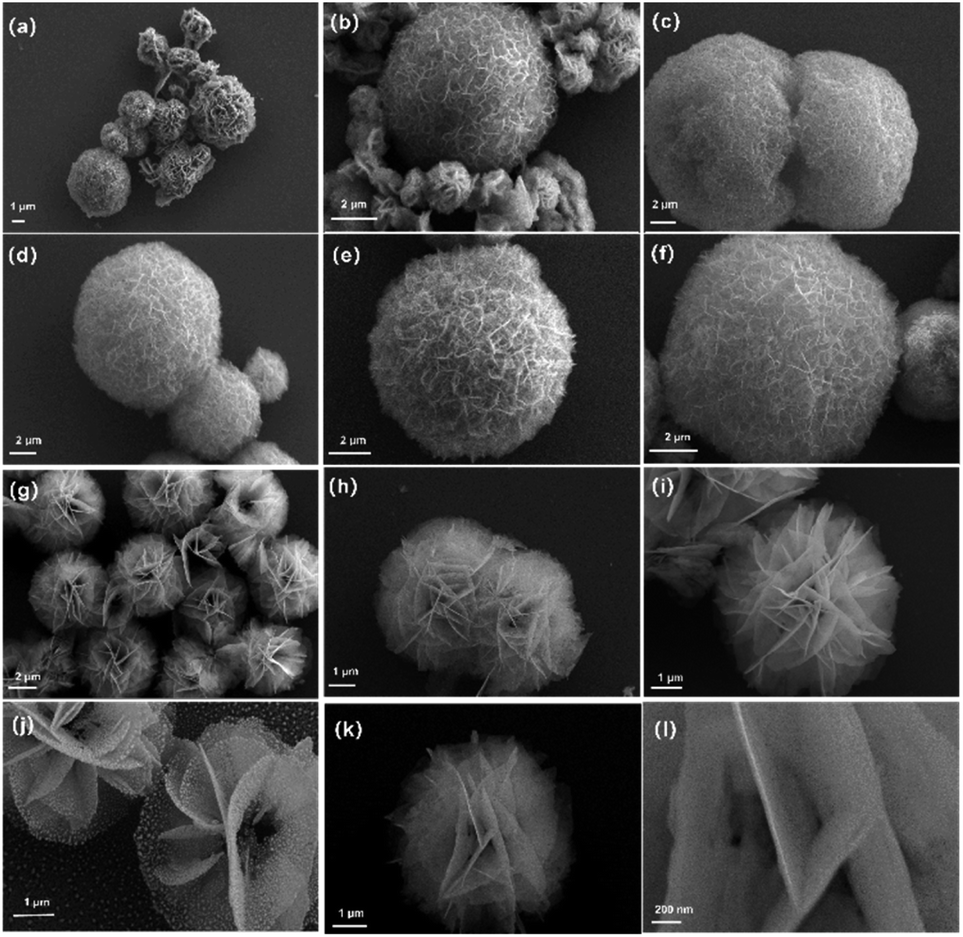
Nanoflower Ni(OH) 2 grown in situ on Ni foam for high-performance  supercapacitor electrode materials - Sustainable Energy & Fuels (RSC  Publishing) DOI:10.1039/D1SE01036K
Nanoflower Ni(OH) 2 grown in situ on Ni foam for high-performance supercapacitor electrode materials - Sustainable Energy & Fuels (RSC Publishing) DOI:10.1039/D1SE01036K
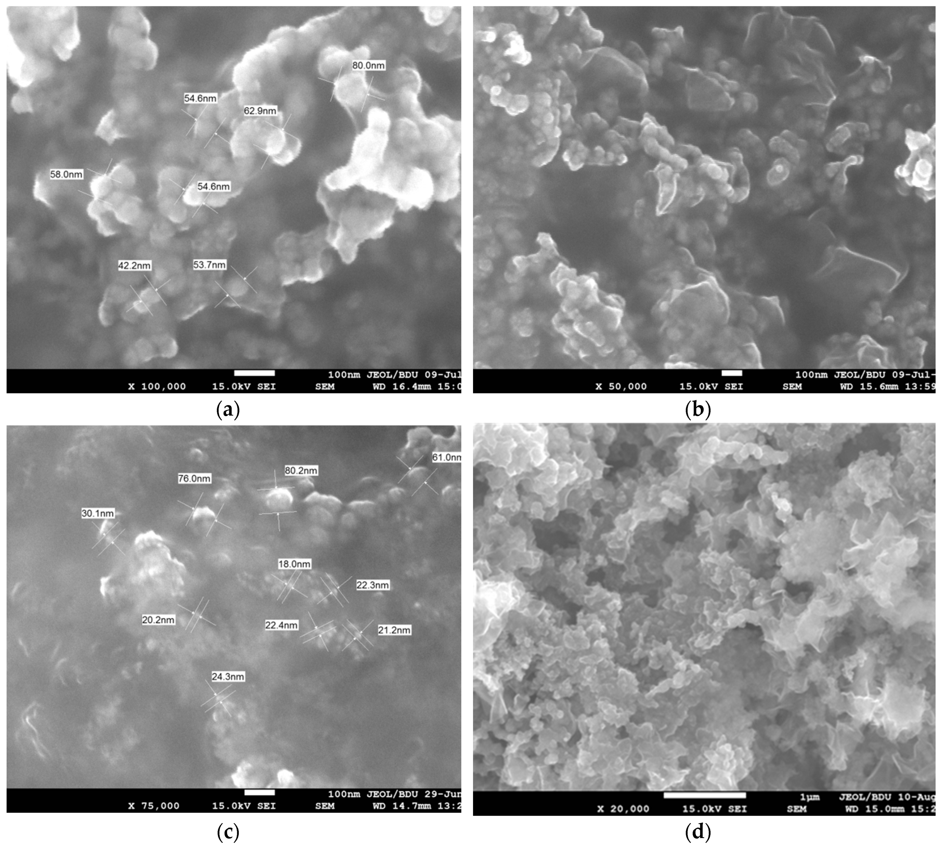
Nanomaterials | Free Full-Text | Synthesis of Fe/Ni Bimetallic  Nanoparticles and Application to the Catalytic Removal of Nitrates from  Water
Nanomaterials | Free Full-Text | Synthesis of Fe/Ni Bimetallic Nanoparticles and Application to the Catalytic Removal of Nitrates from Water

SEM image of synthesized Nickel nanoparticles | Download Scientific Diagram

SEM images of the Ni foam (a), NiO nanosheets on Ni foam (b – d) with... |  Download Scientific Diagram
SEM images of the Ni foam (a), NiO nanosheets on Ni foam (b – d) with... | Download Scientific Diagram

SEM micrograph of a Ni foam obtained at a magnification of 200× | Download  Scientific Diagram
SEM micrograph of a Ni foam obtained at a magnification of 200× | Download Scientific Diagram

SEM images of fresh Ni/zeolites catalyst. (a) Ni/ZSM5-30, (b)... | Download  Scientific Diagram
SEM images of fresh Ni/zeolites catalyst. (a) Ni/ZSM5-30, (b)... | Download Scientific Diagram

Three-dimensional microstructural characterization of solid oxide  electrolysis cell with Ce0.8Gd0.2O2-infiltrated Ni/YSZ electrode using  focused ion beam-scanning electron microscopy | SpringerLink
Three-dimensional microstructural characterization of solid oxide electrolysis cell with Ce0.8Gd0.2O2-infiltrated Ni/YSZ electrode using focused ion beam-scanning electron microscopy | SpringerLink

Nickel Nanoparticles / nanopowder (Ni, 99.5%, 180nm)

SEM and Elemental Mapping study of mechanically alloyed binary W-Ni - 2019  - Wiley Analytical Science
SEM and Elemental Mapping study of mechanically alloyed binary W-Ni - 2019 - Wiley Analytical Science
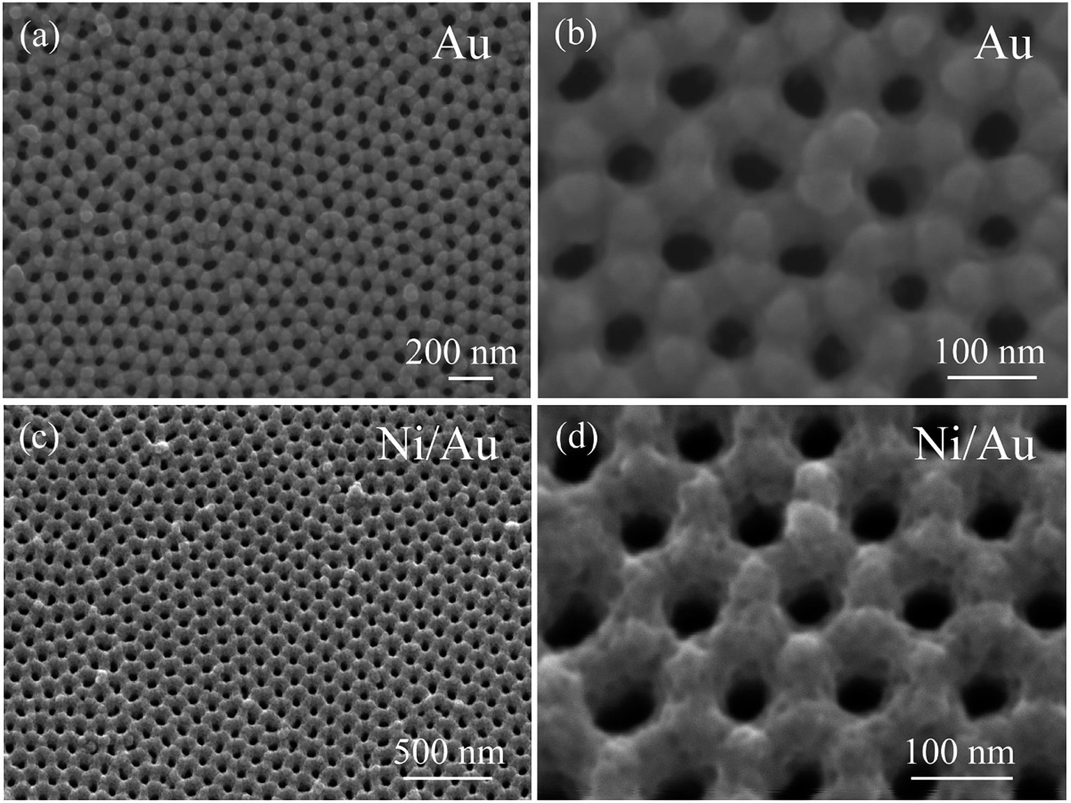
Synthesis of an ordered nanoporous Cu/Ni/Au film for sensitive  non-enzymatic glucose sensing - RSC Advances (RSC Publishing)  DOI:10.1039/D0RA01224F
Synthesis of an ordered nanoporous Cu/Ni/Au film for sensitive non-enzymatic glucose sensing - RSC Advances (RSC Publishing) DOI:10.1039/D0RA01224F

Scanning electron microscopy (SEM) images of Ni-alloyed iron pyrite... |  Download Scientific Diagram
Scanning electron microscopy (SEM) images of Ni-alloyed iron pyrite... | Download Scientific Diagram

SEM images of pure Ni foam (a), and NiFe LDH (b), (c), and (d). | Download  Scientific Diagram
SEM images of pure Ni foam (a), and NiFe LDH (b), (c), and (d). | Download Scientific Diagram